
DI-X1
X-ray Motion Analysis Workstation

In this system, general-purpose radiography equipment can be used, just as for conventional simple chest radiography. This system transmits a series of pulsed X-rays about 15 times per second using the same principle as animation, and displays a series of static images to create dynamic images. Chest dynamic digital radiography images allow us to observe actual movement, and we feel they can provide much more information than static images. In addition, the system allows standing-position radiography, as opposed to supine-position CT or MRI. Therefore, it also has the advantage of being able to observe body conditions in the posture in everyday life.
Radiography enabled in the general radiography environment
Frame rate: 15 fps
Exposure duration: Max. 20 seconds
Dynamic digital radiography - a new tool that offers more information
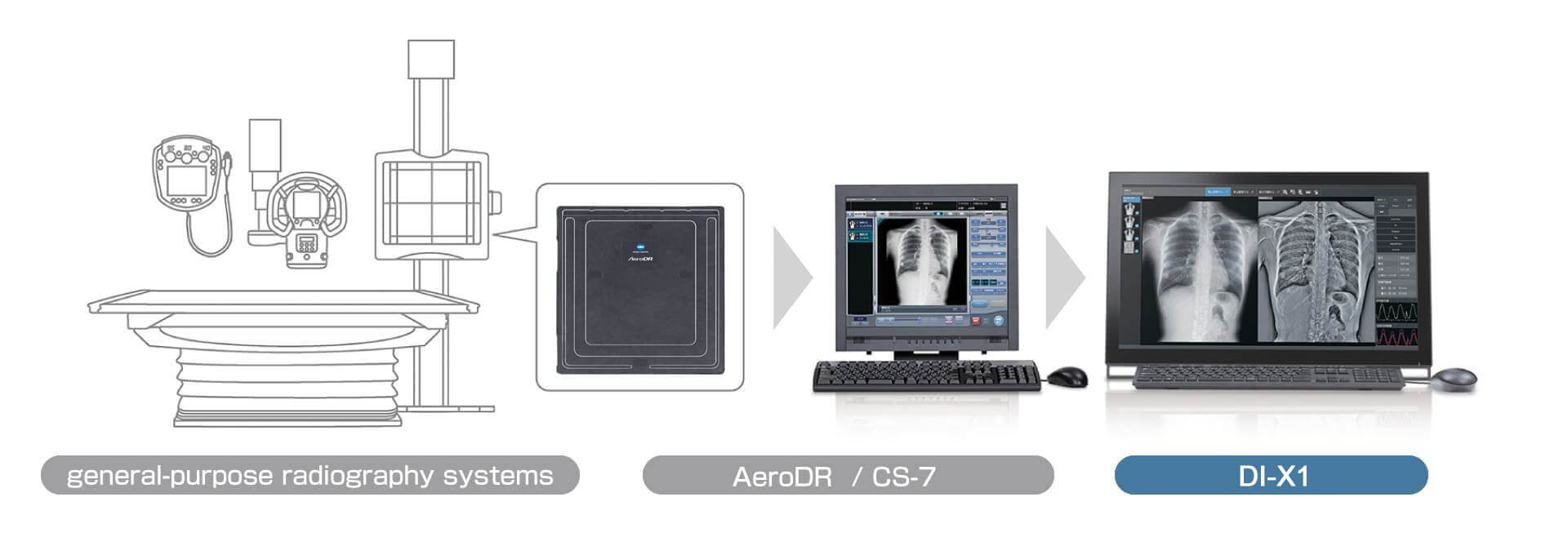
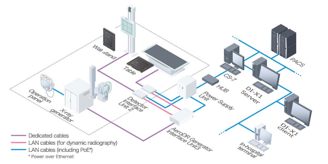
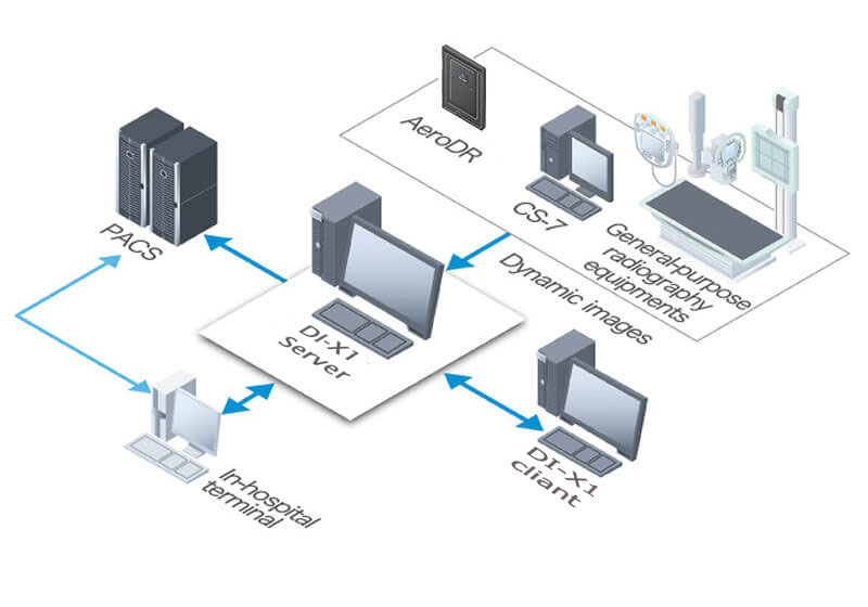
The Dynamic Digital Radiography Analysis Workstation “DI-X1”, the portable DR “AeroDR 3”, and general-purpose radiography systems conventionally used for chest radiography, make up the components of the Dynamic Digital Radiography system. This breakthrough system can transmit a sequence of pulsed X-rays and display a series of static images to create dynamic images. DI-X1 is equipped with Konica Minolta’s proprietary image processing technology to provide a variety of information for the medical field.
Indentification Capability-enhancing Image Processing Technology
The image processing we developed to improve visibility for simple X-ray static images is also applied for the dynamic images.
BS-MODE (Bone Suppression)
By attenuating clavicles and ribs in lung fields to improve visibility, allows us to visualize what was previously difficult to see due to overlap with bone components.
FE-MODE (Frequency Enhancement)
The edges of each structure are made to stand out to improve visibility and facilitate the observation of motion.
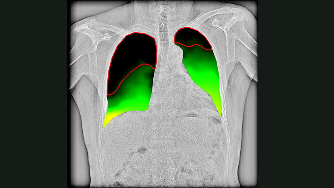
LM-MODE (Option)※
Display in color the amount of movement of each region associated with respiration by tracking the signal values within the lung field.
※option
LM-MODE (Dynamic)※
Added motion vector images as the basis for LM-MODE.
※option
Quantifying "motion"
The motion of structures such as diaphragms can be quantified and displayed graphically. By quantifying motions, the system can provide new perspective to the evaluation of symptoms and functions.
DM-MODE
Tracks the movement of the diaphragm in each frame, measures the amount of vertical movement, and displays it in a graph.
TD-MODE (Chronological order)
The maximum and minimum tracheal diameter in each frame are displayed.
Multipurpose measurement tool (Position change)※
Measure the positional change of a specified, point/line/two line segments. Displays the measurement results in a graph.
※option
Multipurpose measurement tool (Signal value change)※
Measure the signal value change in the specified rectangle/arbitrary area. Displays the measurement results in a graph.
※option
Motion Analysis Technology with the aim of Functional Assessment
The motion of structures such as diaphragms can be quantified and displayed graphically. Motion quantification (graphical display) increases the visibility of the motion itself at a particular point and provides information not previously available from static images for symptom evaluation and functional assessment.
PL-MODE (Reference frame ratio calculation )
Extracts and displays changes in the lung field concentration associated with breathing.
PH-MODE (Cross-correlation calculation)
Extracts and displays changes in the lung field concentration associated with vascular pulsation.
PH2-MODE (Option)※
Extracts and displays the signal value change in the lung field associated with the breathing of blood vessels.
※option
PH2-MODO (Option)※
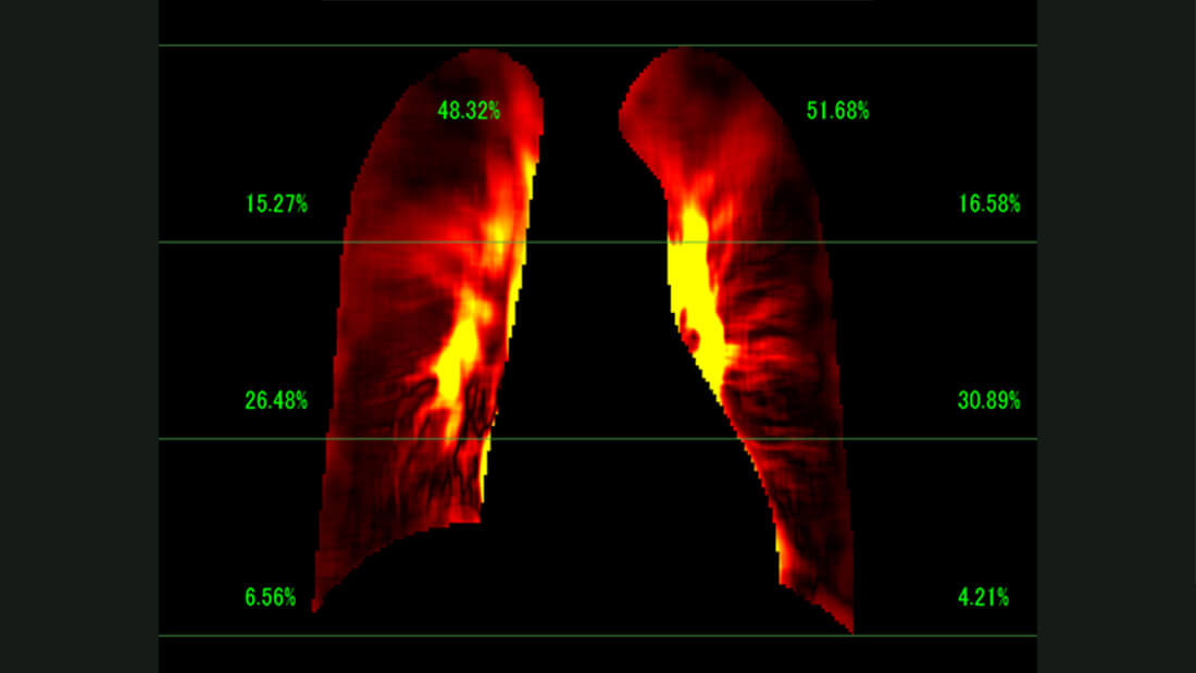
Signal value ratio per lung Region
Calculate the signal value ratio of 6 areas,
※option
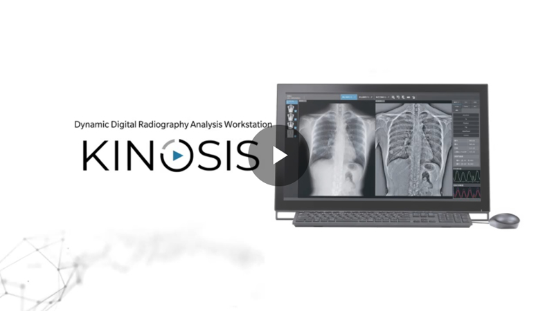
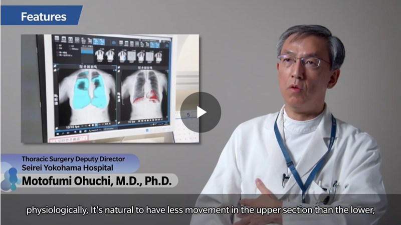
Introductory video: Doctor interviews
The motion of structures such as diaphragms can be quantified and displayed graphically. Motion quantification (graphical display) increases the visibility of the motion itself at a particular point and provides information not previously available from static images for symptom evaluation and functional assessment.

Specifications
System configurations

*This site is intended for healthcare workers.