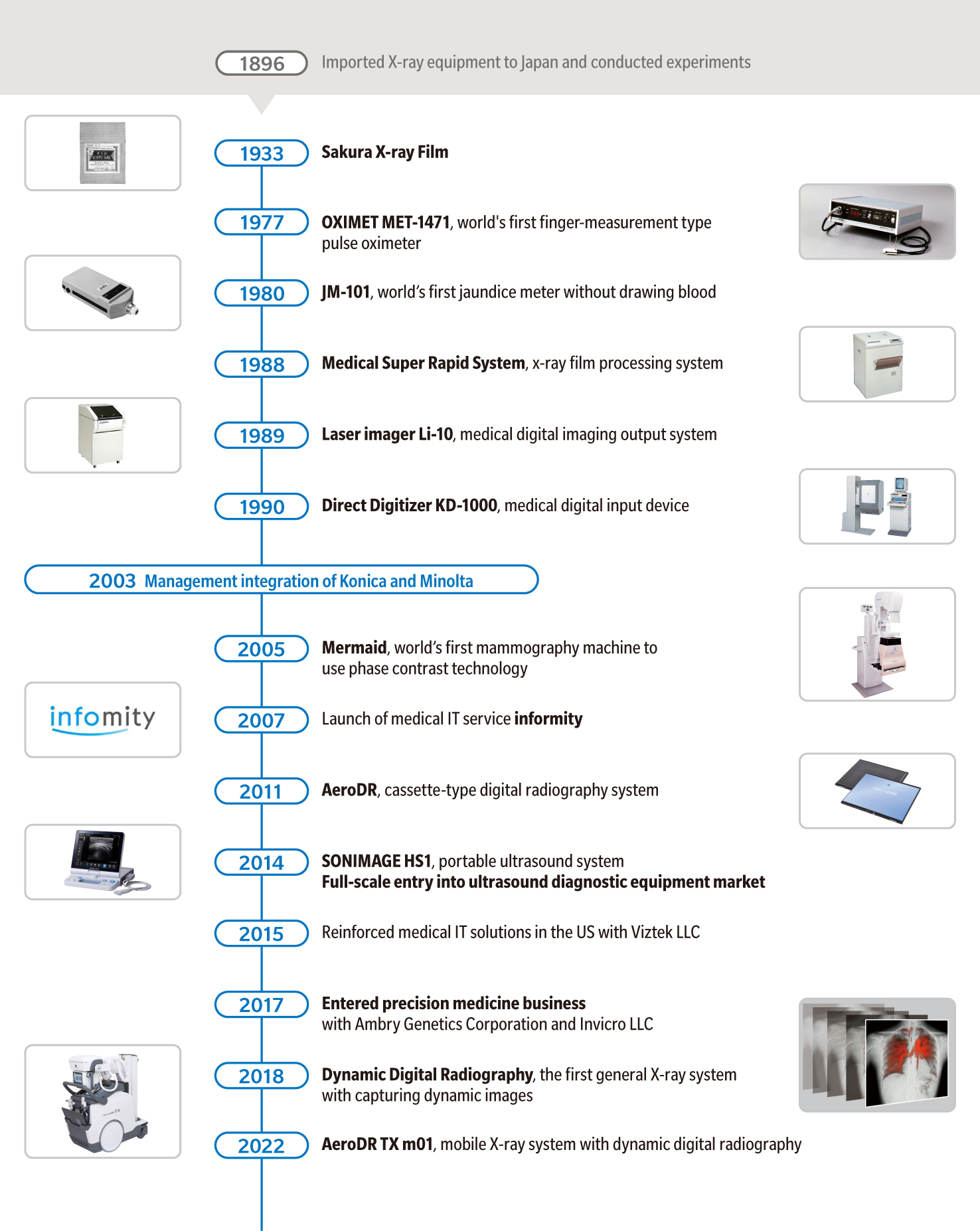
INNOVATION STORIES
HEALTHCARE
Driven by passion to contribute to medicine
History of diagnostic imaging through ongoing co-creation of value with hospitals and clinics
- INDEX
- ・Our desire to contribute to medicine by quickly identifying diseases
- ・Overcoming challenges posed by the major trend of digitalization through co-creation with medical professionals
- ・Entering the ultrasound market to grow the market with hospitals and clinics
- ・Supporting DX of primary care doctors with specialist doctors
X-rays were discovered by a German scientist in 1895. When it was reported that X-rays could take photographs inside the human body, the news spread quickly around the world. We immediately saw the potential of diagnosis using X-rays, leading to the launch of our healthcare business.
Since then, we have expanded our business mainly in the field of diagnostic imaging. Our goals have remained the same: to offer advanced medical diagnosis to as many people as possible, reduce the burden on patients, and improve the workflow at hospitals and clinics. Our healthcare business, which has a history of over 100 years, has developed by passing on the passion and continuously working on co-creation with medical professionals.
Our desire to contribute to medicine by quickly identifying diseases
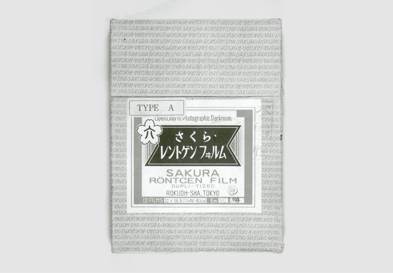
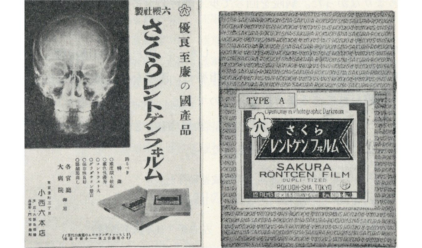
In 1933, Konica released the Sakura X-ray Film, a medical X-ray film which it had developed and produced internally. The world was heading into World War II. Why did the company decide to start producing X-ray films at such a time?
In fact, in 1896, 37 years before the Sakura X-ray Film was launched, our predecessors had
imported X-ray equipment into Japan and conducted various research projects with doctors.
This was just one year after X-rays were discovered by Wilhelm Conrad Röntgen, a German
physicist. Clearly, our predecessors had foresight and the ability to take action.
Our passion for X-ray films was underpinned by a strong desire to contribute to medicine by quickly identifying diseases. In Japan, tuberculosis was rampant from the mid-19th century to the mid-20th century. It was feared as a “national disease.” Our predecessors continued to conduct research, believing that lives could be saved if domestically produced films entered widespread use instead of expensive X-ray films manufactured overseas. Their efforts finally led to the development of the Sakura X-ray Film, which was highly evaluated by doctors and radiographers.
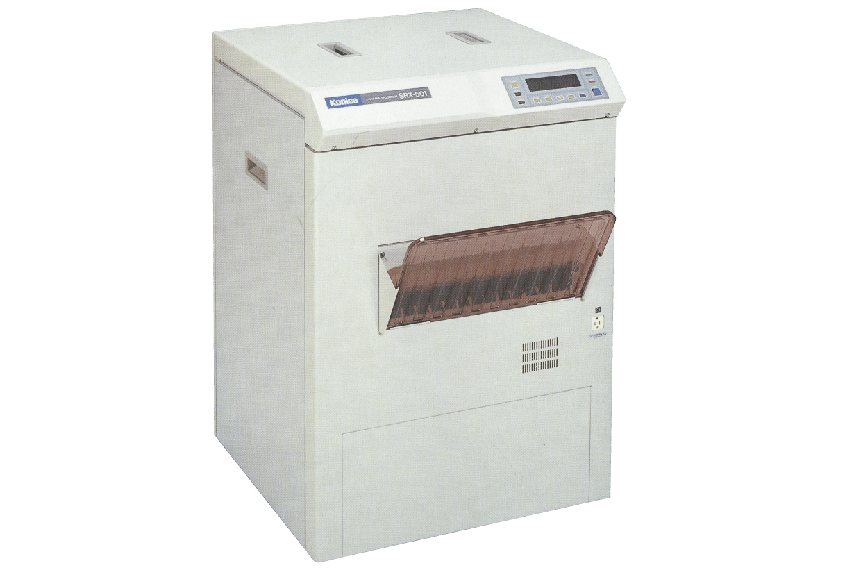
Later, the use of X-ray film increased as X-ray imaging spread at hospitals and clinics. In the 1960s, automatic film developing machines came into use. In 1963, Konica released a product that could develop the film in just eight minutes. Later, the company reduced that development time to 210 seconds, and then to 90 seconds. In 1988, it released the Konica Medical Super Rapid System, a high-speed automatic developing machine that achieved processing in 45 seconds, the fastest in the world. The system was highly evaluated for significantly improving the workflow of emergency medical treatment, in which every second counts.
Overcoming challenges posed by the major trend of digitalization through co-creation with medical professionals
In our long-established healthcare business, the biggest turning point was the shift away from using films in X-ray diagnostic imaging.
In the 1980s, as computers became increasingly used in offices, X-ray diagnostic imaging was left behind, without being digitalized. Diagnostic systems capable of converting X-ray imaging information into digital data and displaying images on a monitor were already on the market, but image quality was considered inferior to film. Nevertheless, there was an unceasing trend toward digitalization of diagnostic imaging due to its many advantages, including the ability to transmit digital images and ease of image processing.

In Japan, digitalization spread rapidly because the digital storage of diagnostic images was permitted following the revision of medical fees in 2008. While the shipment volume of films decreased sharply and the scale of business shrank, we continued to work on digitalization with support from medical professionals, who were our customers.
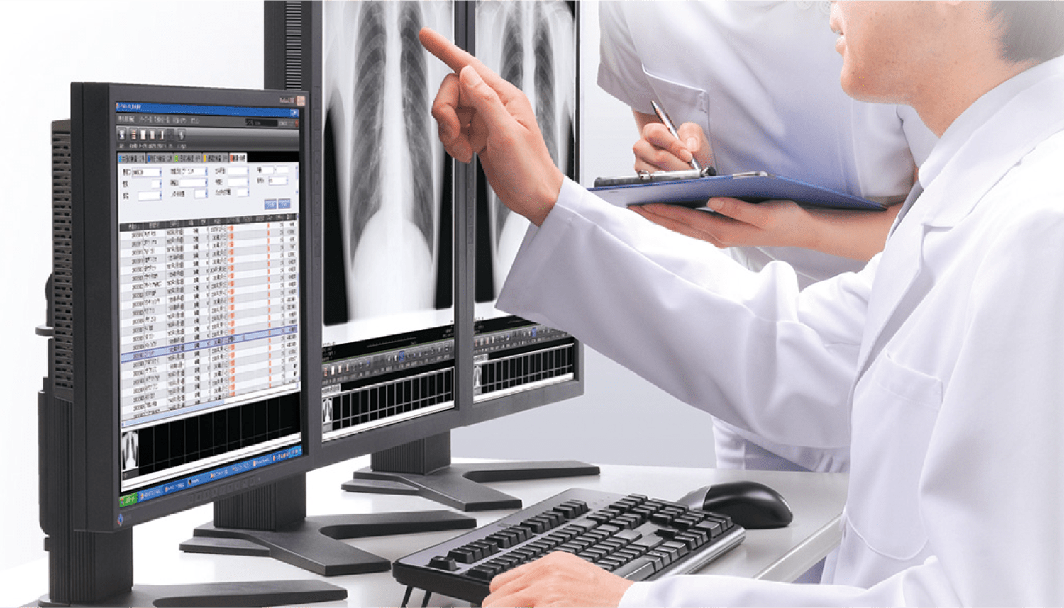
The digitalization of X-ray imaging was an issue for manufacturers. It also posed difficulties for medical professionals because until then they acquired expertise in diagnostic imaging based on the appearance on X-ray films. The person in charge at that time noted, “In diagnostic imaging, doctors identify abnormal conditions, namely, lesions, based on an understanding of images in the normal condition. We studied with doctors how digital images would look differently and how the monitor should be adjusted, and solved the issues one by one.” We overcame this major shift under the heavy expectations of medical professionals.
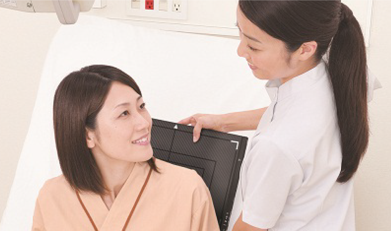
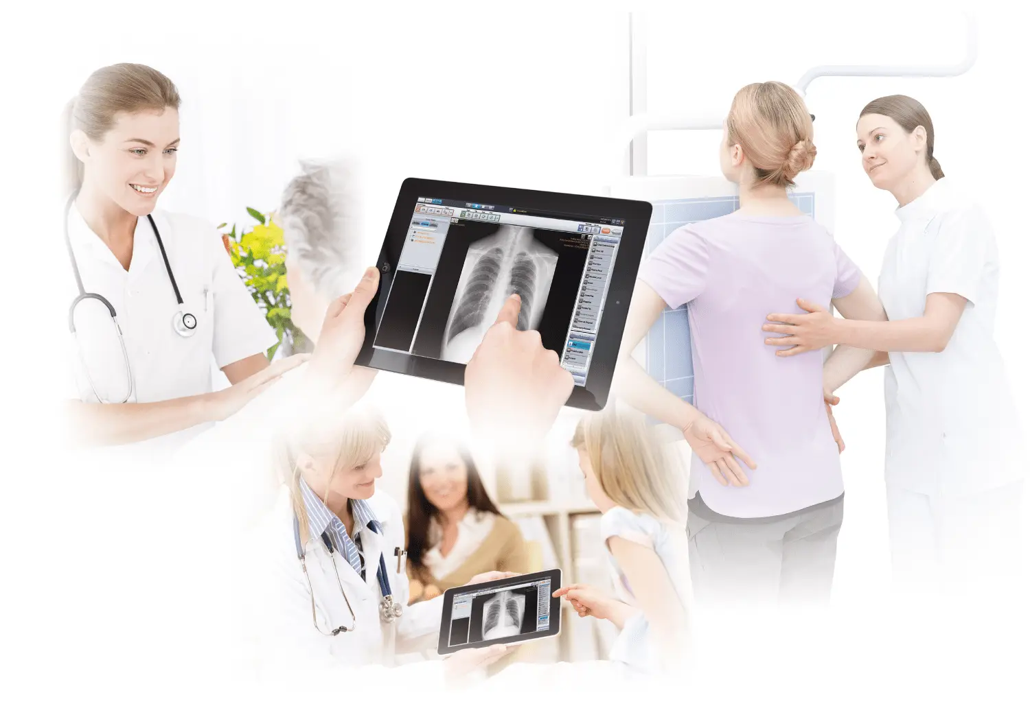
In 2011, we launched AeroDR, the lightest cassette-type digital X-ray system in the world, and built up one of the largest shares in the digital radiography (DR) market in Japan.
* Digital Radiography : X-rays are converted into light by using fluorescent substances. The light is then turned into electric signals by semiconductors, making it possible to display X-ray images on the monitor immediately after taking an X-ray.
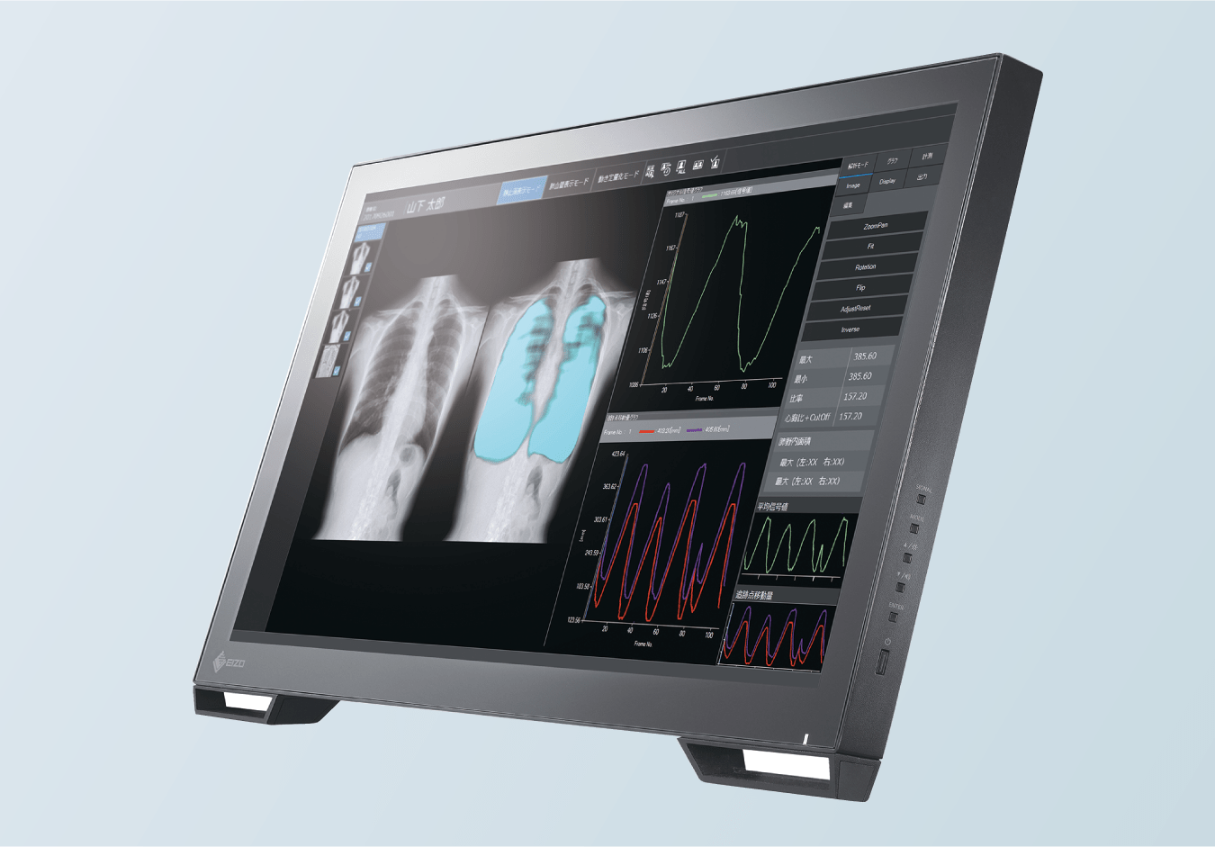
Subsequently, we continued to take on challenges, and in 2018, finally released a product that revolutionized diagnostic imaging: Dynamic Digital Radiography, the world’s first dynamic radiography system using general X-ray imaging equipment. This enables radiography of patients who are unconscious or who cannot hold their breath due to respiratory failure. In combination with our image analysis technologies, we have made it possible to check whether organs are functioning properly at a glance. The system has been highly evaluated by doctors and researchers for bringing about a paradigm shift in X-ray diagnosis, making what was previously invisible visible. The system is increasingly being used in many countries around the world.
Entering the ultrasound market to grow the market with hospitals and clinics
Following the digitalization of X-ray imaging, our initiative to develop diagnostic ultrasound systems marked the next pivotal step in the healthcare business.
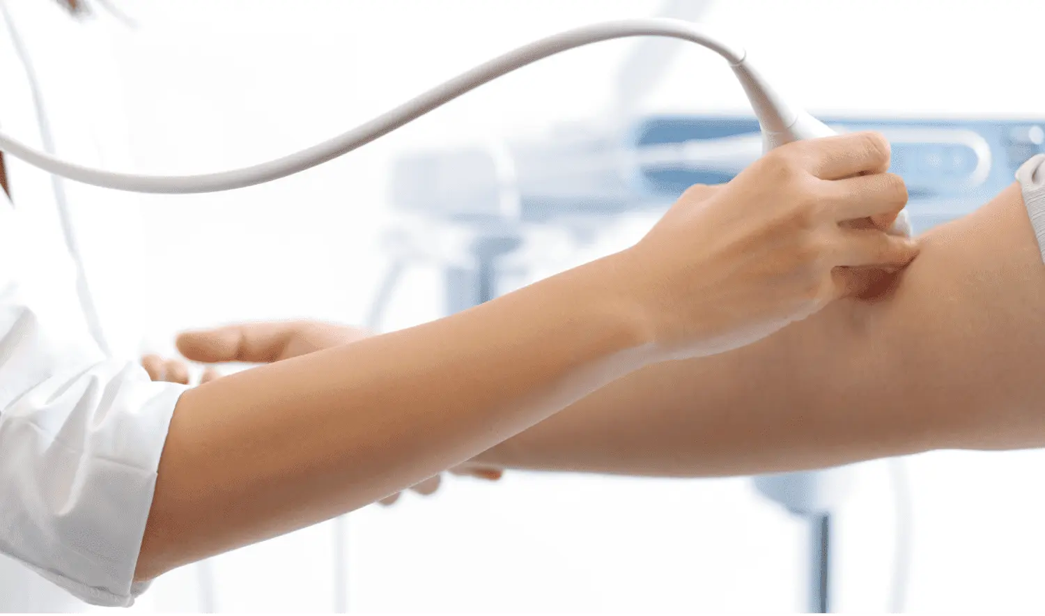
Diagnostic ultrasound (echo) systems utilize sound waves, which are inaudible to the human ear, to perform diagnosis. They can be used in various diagnostic fields because the physical burden on patients is minimal and images can be checked in real time. We entered this field on a full scale in 2014 with the release of the SONIMAGE HS1, a product jointly developed with the ultrasound division of Panasonic Healthcare Co., Ltd., which an entity we had previously integrated.
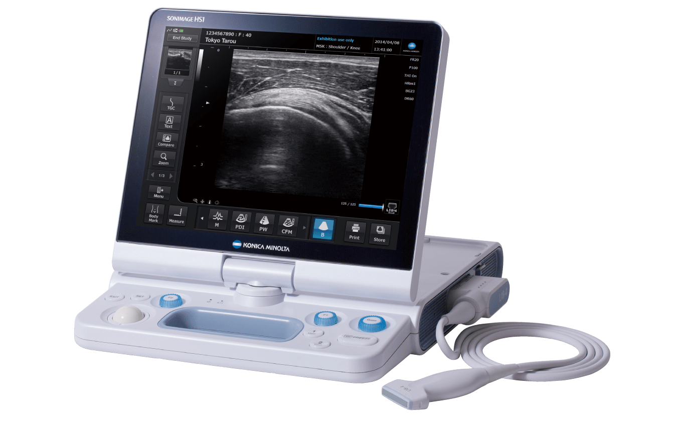
We thought we could make ground-breaking technological innovations in probes, crucial components placed on the patient’s body surface. Probes emit ultrasound waves and receives echoes, and determines the resolution of a system. In developing probes, we leveraged our material technology cultivated through film development, and our image processing technology cultivated in X-ray diagnostic imaging. We succeeded in developing equipment that produces high-quality images that clearly show the fiber structure of muscle fascicles and nerve fascicles, which are just several tens to hundreds of micrometers thick.
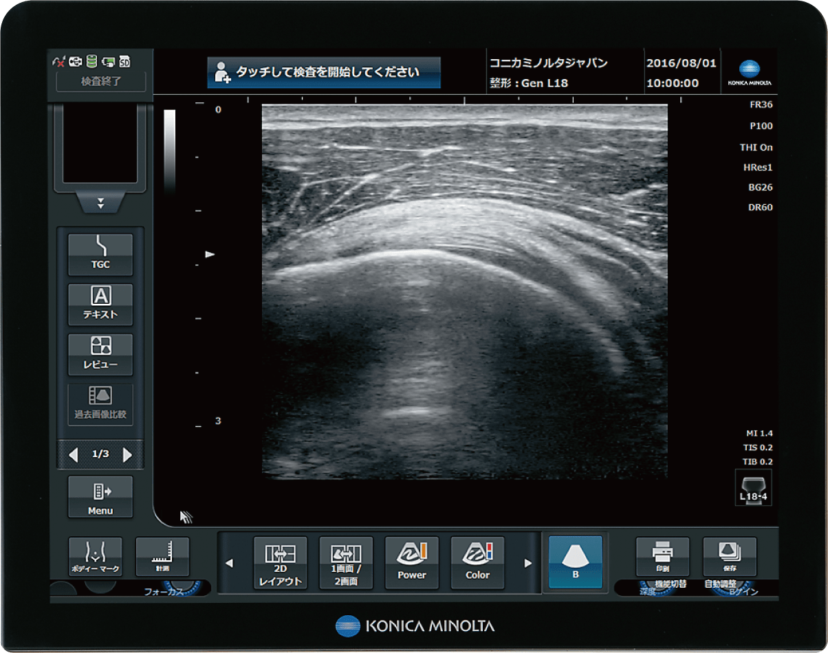
At that time, we were the last manufacturer to enter the diagnostic ultrasound market. We had no market share when we started, so we decided to start with orthopedics, where we thought we could take advantage of our strengths. We conducted various tests with doctors, who were key opinion leaders, to identify the field to which we could contribute most.
Another focus in product development was user friendliness. The first reaction from doctors when we showed our prototype was harsh comments to improve user friendliness, such as “This button is unnecessary,” and “This is difficult to use.” We were confident about the performance of the equipment, including the probe, so we carefully listened to the feedback from users at hospitals and clinics about the button positions, screen configuration, and operation method, and reflected that feedback in product development.
As a result, about two years after its release, the SONIMAGE HS1 secured the top share in the orthopedics field. Later, the number of doctors starting to use echo for diagnosis increased through seminars and other events, and the number of units sold grew as we worked to open up the market. Today, our diagnostic ultrasound systems are also used in other fields. Our constant communication with hospitals and clinics has enabled us to win the trust of many medical professionals, receive candid opinions, and grow the market together.
Supporting DX of primary care doctors with specialist doctors
We have begun to support digital transformation (DX) of medicine with ICT. This is achieved by a cloud service based on “infomity,” our medical ICT platform that has been introduced mainly to clinics in Japan.
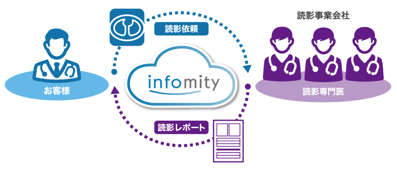
Launched in 2007, the service has been performing stably for 16 years based on a 24/7 call center and the cloud monitoring system, which give customers a sense of security. Both fixed and variable fee services are available. The introduction of a subscription business model in medical services was new at that time. At first, we offered a 24/7 remote maintenance service and an image storage service. Recently, we offer the Collaboration Box Service, which enables multiple medical institutions to share examination information, including imaging data, a teleradiology support service, which enables primary care doctors to ask specialist doctors interpretation, and an image analysis service using artificial intelligence (AI). In 2023, we started to offer the “infomity smart clinic service” to facilitate information linkage and communication by connecting patients’ smartphones with hospitals and clinics.
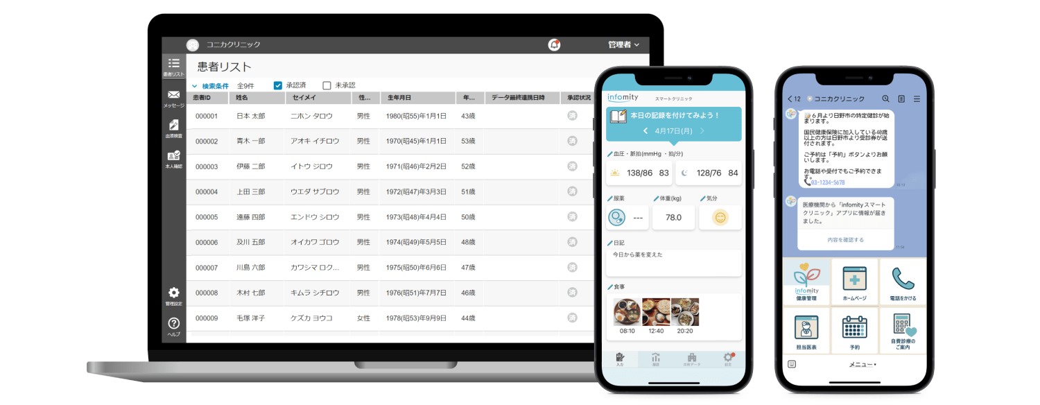
In Japan, there are more than 100,000 general clinics, and X-ray diagnostic imaging is performed at about half of them. Of these, about 20,000 clinics (40%) are connected with infomity, which serves as an inexpensive and convenient tool and as a key DX solution for primary care doctors, who are close to patients. We are continuing to improve our systems through collaboration with many specialist doctors to implement cutting-edge diagnosis support technologies in infomity.
For more than a century, Konica Minolta’s healthcare business has gone through many turning points. However, we remain committed to saving lives whenever possible and co-creating value with medical professionals. Our DNA will continue to help improve workflow efficiency at medical institutions and support healthy, high-quality living.
In the interest of clarity, “Konica” and “Minolta,”—the trade names that were used before the merger that formed “Konica Minolta”—are used throughout this text. Each company underwent a number of name changes throughout their history.