
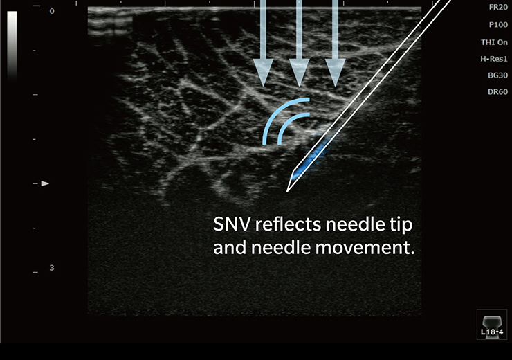


SNV
Simple Needle Visualization
- Available for both in-plane and out-of-plane approaches in the ultrasound-guided procedure
- Achieves "steep needling" visualization
- Keeps the original B-mode image quality
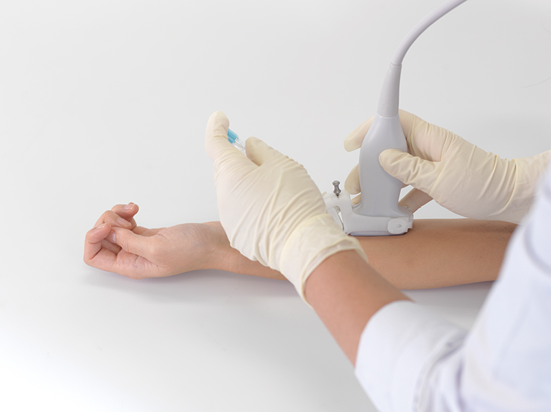
Available for both in-plane and out-of-plane approaches in the ultrasound-guided procedure
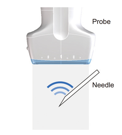
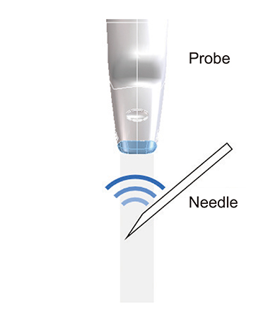
Generally, the ultrasound-guided procedure has two needling approaches. One is in-plane approach where needle moves parallelly to the image plane. The other approach is out-of-plane where it moves across the image plane.
In in-plane approach, especially when it comes to the steep needling, sensitivity of the needle in B-mode images becomes weaker due to lack of reflection signals from it. In out-of-plane approach, it is difficult to find the timing when needle passes through the image plane.
Our SNV is available for both the approaches by introducing our original technique.
Achieves "steep needling" visualization
As described above, sensitivity of needle becomes weaker especially in steep needling cases. To compensate that, our SNV introduced the multi-frame analysis to detect needle motion. The needle motion helps enhance needle sensitivity especially in the case of steep needling. Through this technique, SNV achieves the "steep needling" visualization.
Keeps the original B-mode image quality
B-mode image quality is highly required for safety needling. The conventional visualization based on the ultrasound beam steering degrade original B-mode image with its artifacts and it also limits range of the field of view.
Our SNV can detect needle motion by introducing multi-frames analysis. This technique allows us to keep the original B-mode image quality and also visualize the needle. Users are not interfered with any artifacts and range limitation of the field of view.
Ultrasound technology
Digital Radiography technology
*This site is intended for healthcare workers.